A brain haemorrhage is defined as a bleed in the brain tissue and it is the third leading cause of mortality across all ages. Brain Haemorrhages are caused either by a haemorrhagic stroke, or a significant blow to the head [1]. A brain haemorrhage is often treated as an emergency since it can lead to death, hence the diagnosis process is very time-critical. One of the most commonly used diagnostic tools for patients being treated for a brain injury or patients with symptoms of a stroke or rise in the intracranial pressure is a non-contrast Computed Tomography (CT) scan or a Magnetic Resonance Imaging (MRI) scan [2].
Computer-Aided Diagnosis (CAD) systems have been developed and were introduced to aid radiologists and professionals in their decision making. CAD systems are intended to be used as a tool to aid radiologists, rather than replace them. Deep Learning CAD systems were not highly researched before, but due to recent advancements in technology, deep learning algorithms have become more popular, and are now being researched for their applications in medical imaging [3].
This study utilizes deep learning models to develop a computer aided diagnosis (CAD) system to classify the different types of haemorrhages in head CT scans. The system was designed in such a way that it builds upon the work done on the final year projects of Mr. John Napier and Ms. Kirsty Sant. Mr. Napier’s work focused on developing a system that can detect the presence of a brain haemorrhage in CT scans, and Ms. Sant’s work involved using a machine learning technique to classify the brain haemorrhages, based on the intensity, shape and texture features [4], [5].
Deep learning architectures, namely ResNet, DenseNet, and InceptionV3 architectures, were analysed in order to find the best performing architecture to classify the different types of brain haemorrhages from head CT scans. Moreover, a linear Support Vector Machine was also built in order to be able to compare the performance of these architectures with it.
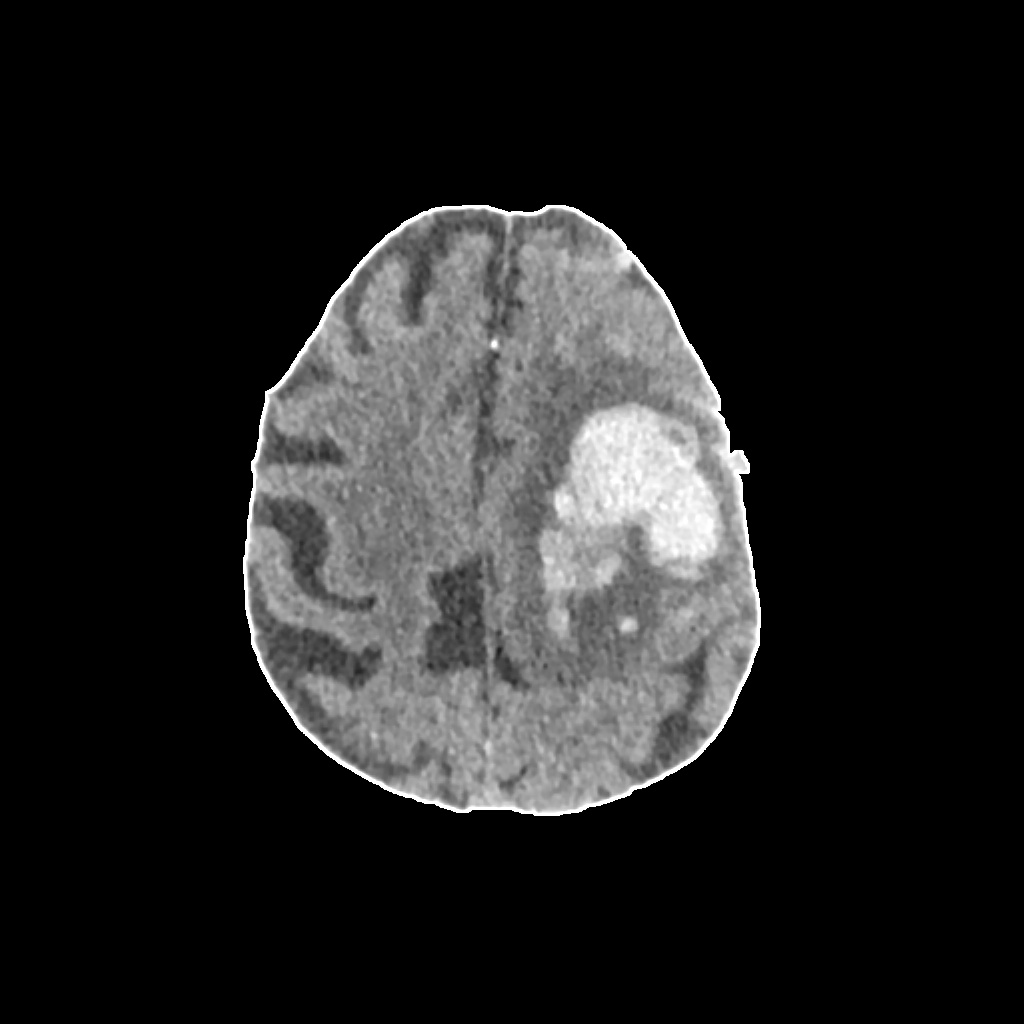
The dataset was obtained from the General Hospital of Malta and contained 64 anonymised brain haemorrhage CT scans. 58 of these were used for training the deep learning models, while the remaining 6 cases were used to test the models. Each of the architectures was executed for 100 epochs, and the overall training accuracy was 0.1786 for ResNet, 0.2976 for DenseNet, 0.3690 for InceptionV3 and 0.6083 for the linear multiclass support vector machine.
References
[1] M. I. Aguilar and T. G. Brott, “Update in intracerebral haemorrhage,” The Neurohospitalist, vol. 1, (3), pp. 148-159, 07, 2011.
[2] Chilamkurthy et al, “Development and validation of deep learning algorithms for detection of critical findings in head CT scans,” 12 April. 2018.
[3] K. Doi, “Computer-aided diagnosis in medical imaging: historical review, current status and future potential,” Computerized Medical Imaging and Graphics : The Official Journal of the Computerized Medical Imaging Society, vol. 31, (4-5), pp. 198-211, 2007.
[4] J. Napier, “Image processing techniques for brain haemorrhage detection in head CT scans,” 2017. [5] K. Sant, “Classification of brain haemorrhages in head CT scans,” 2018.
Student: Nicola’ Spiteri
Supervisor: Prof. Inġ. Carl James Debono
Co-supervisors: Dr Paul Bezzina and Dr Francis Zarb
Course: B.Sc. (Hons.) Computer Engineering